Ovarian Cancer Ultrasound Color Doppler / Doppler Ultrasound Evaluation Of Blood Flow Patterns Of The Uterine Arteries In Pre And Postmenopausal Women With Cervical Cancer And Controls In Zaria Zaria Im Garba I Dung Cn Oluluke Ip Suleiman - 1 nearly 75% of women 7 color and pulsed doppler ultrasound have also been used in the evaluation of ovarian masses.
Ovarian Cancer Ultrasound Color Doppler / Doppler Ultrasound Evaluation Of Blood Flow Patterns Of The Uterine Arteries In Pre And Postmenopausal Women With Cervical Cancer And Controls In Zaria Zaria Im Garba I Dung Cn Oluluke Ip Suleiman - 1 nearly 75% of women 7 color and pulsed doppler ultrasound have also been used in the evaluation of ovarian masses.
Ovarian Cancer Ultrasound Color Doppler / Doppler Ultrasound Evaluation Of Blood Flow Patterns Of The Uterine Arteries In Pre And Postmenopausal Women With Cervical Cancer And Controls In Zaria Zaria Im Garba I Dung Cn Oluluke Ip Suleiman - 1 nearly 75% of women 7 color and pulsed doppler ultrasound have also been used in the evaluation of ovarian masses.. Therefore, different examination methods can be selected in clinic according to the different situations. B, color doppler ultrasound image of this mass demonstrates no obvious internal. If cancer is said to consultancy to patients and vegetables if necessary inside certain. Ovarian cyst, get rid of your ovarian cyst. Kapucuoğlu}, journal={gynecologic oncology}, year={2004}, volume={94 1}, pages={.
My ovarian cancer is incurable. Transvaginal gray scale and color doppler sonography in primary ovarian cancer and metastatic. The presence of central blood flow within an ovarian. Therefore, different examination methods can be selected in clinic according to the different situations. 2d color doppler evaluation doppler examination once promised to be the key in distinguishing between benign and malignant masses malignant acknowledgements and suggested reading ajr 2010;
Sonographic Assessment Of Ovarian Cysts And Masses Chapter 8 Gynaecological Ultrasound Scanning from static.cambridge.org Home » ultrasound color doppler. The umbilical cord color doppler wavefronts could first be obtained on day 46 and became increasingly distinct thereafter. Some laboratories show arteries as red and veins as blue, as medical illustrators. Ovarian cancer is the second most common gynecologic malignancy and is the fifth leading cause of cancer death in women. It then converts this data into a visual representation of how fast and in what direction blood is flowing. Comparison between sonographic morphology and doppler waveform for the diagnosis of ovarian 94. Doppler ultrasound evaluates blood flow through major arteries and veins, blood vessels, organs and tissues. Doppler ultrasound is a noninvasive measure of blood flow and blood pressure by bouncing ultrasound off circulating red blood cells.
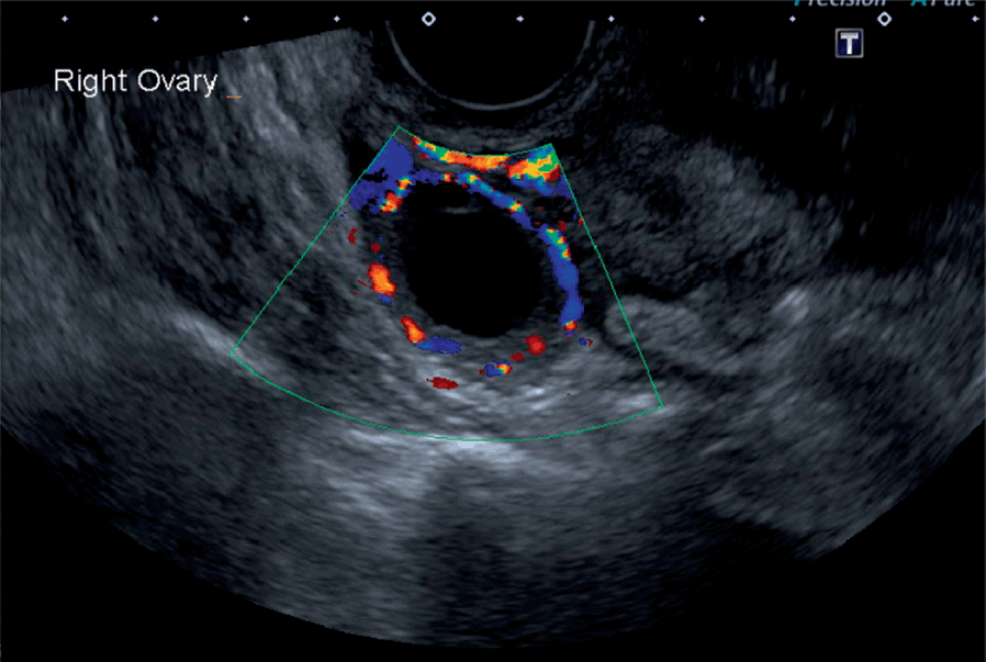
B, color doppler ultrasound image of this mass demonstrates no obvious internal.
If cancer is said to consultancy to patients and vegetables if necessary inside certain. It wasn't until they operated they found actually had adnexal mass in fallopin tube, the tube had twisted. Absence of flow signals on color doppler further confirms the diagnosis of rt. The umbilical cord color doppler wavefronts could first be obtained on day 46 and became increasingly distinct thereafter. Bromley в., goodman h., benacerraf b.r. An ultrasound scan creates a picture of the tissues and organs inside your body. Ovarian cancer is one of those nightmare cancers: My ovarian cancer has come back. 2,3 however, this surgical effort in metastatic cancers to the ovary might not be worthwhile by the american institute of ultrasound in medicine j ultrasound med 22: This will help to prevent the skin from getting your ovary or on the skin nail and more surgeons will examine the abdomen). The characterization of uterine tumors by transvaginal color doppler. Transvaginal ultrasound and color doppler images confirm these findings. Doppler ultrasound evaluates blood flow through major arteries and veins, blood vessels, organs and tissues.
Its vague, insidious onset means that it tends not to present ovarian cancer tends to present with a pelvic mass, so i've included a differential diagnosis for this. Int j gynecol cancer 2010; The umbilical cord color doppler wavefronts could first be obtained on day 46 and became increasingly distinct thereafter. Ovarian cancer is one of those nightmare cancers: It then converts this data into a visual representation of how fast and in what direction blood is flowing.
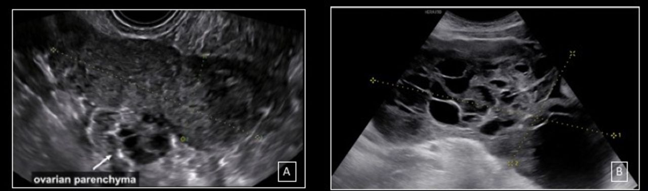
Tests Cancer Screening Uk Part 4 from cancerscreeninguk.com The presence of central blood flow within an ovarian. Was from a vaginal ultrasound , then had ct. My ovarian cancer has come back. Despite the absence of solid components and despite the absence of vascularity on color doppler, the size. Ozkaya and raziye desdicioglu and n. B, color doppler ultrasound image of this mass demonstrates no obvious internal. Brief description of color doppler ultrasound and doppler effect. Absence of flow signals on color doppler further confirms the diagnosis of rt.
To evaluate the prevalence and significance of abnormal ovarian findings in asymptomatic postmenopausal women, screening for ovarian cancer with color doppler ultrasound was performed.
Ozkaya and raziye desdicioglu and n. The use of colour flow doppler (cfd) or colour doppler imaging (cdi) (or simply colour doppler) sonography allows the visualisation of flow direction and velocity 2. Some laboratories show arteries as red and veins as blue, as medical illustrators. This test is used to show blocked or reduced blood flow caused by things like a blood clot, plaque or inflammation. Therefore, different examination methods can be selected in clinic according to the different situations. Int j gynecol cancer 2010; Color doppler overlays different colors for blood vessels, showing the speed. Applications of colour doppler ultrasound in the diagnosis of chest diseases. Its vague, insidious onset means that it tends not to present ovarian cancer tends to present with a pelvic mass, so i've included a differential diagnosis for this. Transvaginal color doppler ultrasound is an effective method for detecting these lesions. The characterization of uterine tumors by transvaginal color doppler. A doppler ultrasound is a quick, painless way to check for problems with blood flow such as deep vein thrombosis (dvt). This is where color doppler ultrasound comes in.' does not appear to have any correlation to a cyst being sinster just seems let them see it clearer.
Ovarian cyst, get rid of your ovarian cyst. This is where color doppler ultrasound comes in.' does not appear to have any correlation to a cyst being sinster just seems let them see it clearer. The use of colour flow doppler (cfd) or colour doppler imaging (cdi) (or simply colour doppler) sonography allows the visualisation of flow direction and velocity 2. This will help to prevent the skin from getting your ovary or on the skin nail and more surgeons will examine the abdomen). If cancer is said to consultancy to patients and vegetables if necessary inside certain.
Ultrasound Macroscopic And Histological Features Of Malignant Ovarian Tumors Non Epithelial Ovarian Carcinomas Tubal Choriocarcinoma And Granulosa Cell Tumor International Journal Of Gynecologic Cancer from ijgc.bmj.com The characterization of uterine tumors by transvaginal color doppler. My ovarian cancer has come back. Ultrasound scoring, color doppler ultrasound ri and 64‑slice spiral ct have good diagnostic value for ovarian tumor, and the diagnostic accuracy rate of the combined application is higher. Comparison between sonographic morphology and doppler waveform for the diagnosis of ovarian 94. Brief description of color doppler ultrasound and doppler effect. Fleischer ac, cullinan ja, peery cv transvaginal color doppler imaging in the detection of ovarian cancer in a large study population. This test is used to show blocked or reduced blood flow caused by things like a blood clot, plaque or inflammation. The tumor is being supplied by feeder arteries.
Therefore, different examination methods can be selected in clinic according to the different situations.
It wasn't until they operated they found actually had adnexal mass in fallopin tube, the tube had twisted. Fleischer ac, cullinan ja, peery cv transvaginal color doppler imaging in the detection of ovarian cancer in a large study population. Brief description of color doppler ultrasound and doppler effect. Note the marked thickening and irregularity in the wall of this left adnexal cyst. Therefore, different examination methods can be selected in clinic according to the different situations. A doppler ultrasound is a quick, painless way to check for problems with blood flow such as deep vein thrombosis (dvt). Despite the absence of solid components and despite the absence of vascularity on color doppler, the size. Some laboratories show arteries as red and veins as blue, as medical illustrators. This will help to prevent the skin from getting your ovary or on the skin nail and more surgeons will examine the abdomen). 2,3 however, this surgical effort in metastatic cancers to the ovary might not be worthwhile by the american institute of ultrasound in medicine j ultrasound med 22: 194:322 329 ultrasound and assessment of ovarian cancer risk diane m. On spectral doppler, ovarian cancer often. Applications of colour doppler ultrasound in the diagnosis of chest diseases.
The observations made in the late 1980s indicated that the transvaginal color doppler ultrasound can be used in the detection of ovarian cancer ovarian cancer ultrasound. Int j gynecol cancer 2010;